Image Submission
This page covers the requirements for submitting an x-ray image for CHEDS.
- the minimum age of a dog for submission under the scheme is one year. There is no upper age limit
- the dog must be permanently identified by microchip and this must be verified by the veterinary surgeon taking the image and placed on the image
- the relevant submission documents must be completed (ie form for either Dogs Australia registered dog, non DA registered dog or dog registered with an overseas kennel club – these forms are atached or can be found on the Dogs Australia website – please dispose of any out of date forms you have) prior to radiography of the dog’s hips, verifying that the details are correct and granting permission for the results to be used in the ways specified.
- The owner must read the declaration and acceptance of terms and confirm agreement.
- Radiographs must be taken under general anaesthesia or heavy sedation
-
Only DICOM images (.dcm) will be read and scored. JPEG or any other images will not be
read (implementation 1 January 2024).
- JPEGS are “snapshots” and usually do not contain enough information for correct identification, scoring and measuring, and will be rejected.
- DICOMs can be large files, and can be difficult to email directly as an atachment to those readers that don’t have a portal. They can be compressed (zipped) before emailing (right click on the file and select“compress”). By doing this, they should be able to be sent as atachments in a single email. Otherwise, the images can be sent one by one as atachments in separate emails.
- Alternatively, file sharing software such as Dropbox, WeTransfer, Google Drive or Microsoft OneDrive can be used to upload the DICOM files as they are. A link is then emailed to the reader, allowing the reader to download them.
- Some digital radiography systems enable the user to email a link which allows the images to be viewed directly from the PACS, thus not requiring the extra steps of transfer or atachment of the individual files themselves. The Metron system and Asteris Keystone Omni are two examples of systems that have this capability.
- Once a certificate of hip and/or elbow scoring has been issued for a dog, the dog’s radiographs may not be resubmited for scoring under the scheme other than via an appeal.
- If the veterinarian taking the images is undecided as to the best image to send, they can send a selection, but no more than three images per required view, always considering the total file sizes to be sent.
Radiographic Technique
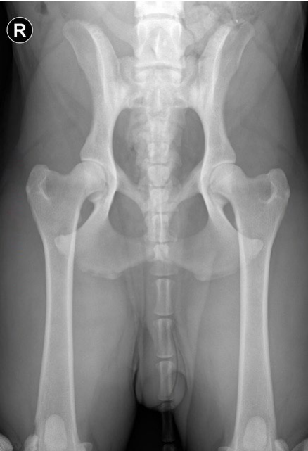
Hips
- A single extended VD view of the pelvis is required for scoring, taken under general anaesthetic or heavy sedation, to enable correct positioning.
- The dog is positioned in dorsal recumbency and the hind limbs extended caudally
- The image should include the wings of the ilia cranially, and if possible, include the stifles (if the size of the dog allows). Priority should be given to including the wings of the ilia rather than the stifles if the dog is too large.
- The x-ray beam is centred over the hips, this can be achieved by palpating bony landmarks such as the cranial edge of the pubic symphysis, and the greater trochanters.
- The femurs are held parallel by rotating the limbs medially, so that the patellas are superimposed over the distal femurs, and adducting the limbs.
- It is important that there is no tilting or rotation of the pelvis, as this can make one hip look beter and the other look worse than it actually is, affecting the scoring. This can be assessed by checking the wings of the ilia, which should look identical; if there is tilting, one iliac wing will look wider than the other, the wider one being tilted down. Also, the obturator foramina should look identical. A well-positioned pelvis looks symmetrical.
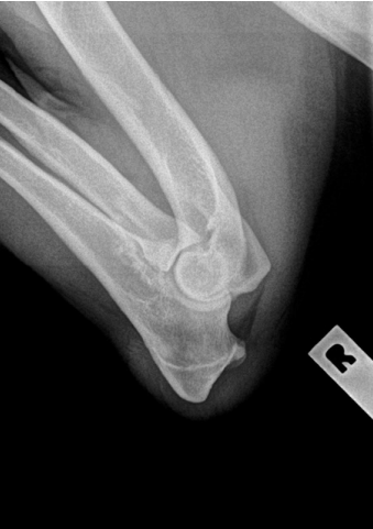
Elbows
- A single, fully flexed, medial to lateral view of each elbow is required, labelled L and R. This allows clear visualisation of the anconeal process, to check for any evidence of osteophyte formation.
- With the dog in lateral recumbency, the elbow to be radiographed is pulled away from the body, to prevent superimposition of the sternum, and then completely flexed. The chest is rolled away from the elbow, and the upper forelimb is pulled caudally.
- The x-ray beam is centred on the elbow joint, this can be achieved by palpating the humeral condyles and using them as landmarks for centring the beam.